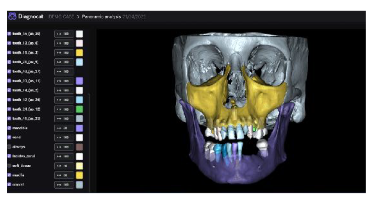
3D imaging is a type of imaging that uses a three-dimensional approach to view the inside of a patient's mouth. 3D imaging renders cross-sectional views of a tooth, which aids in identifying dental problems.
Cone beam computed tomography, or CBCT for short, is a specialized machine that creates a 3D model of the patient's tooth and surrounding structures. The scan is done in seconds and produces for the dentist a three-dimensional image that they can rotate and manipulate to get a better look at any areas of concern. Much like a traditional CT scan that takes images of bone structure, the CBCT scan is non-invasive and provides a wealth of valuable information to your dentist.
Once the scan is complete, our dentist will be able to view a comprehensive view of the teeth and the surrounding tissues. They can even zoom in on specific areas to get a closer look than is possible with a traditional 2-D X-ray. The CBCT technology has revolutionized dentistry by giving dentists more treatment options and information than ever before.
Benefits of 3D Imaging in Dentistry
Your dentist can use a 3D imaging system to scan your teeth, mouth, and gums for problems like tooth decay, gaps between teeth, impacted wisdom teeth, or periodontal disease. The scanner creates a 3D digital model of your entire mouth that our dentist can use to create a customized treatment plan for you.
Our dentist uses 3D imaging for treatment planning in a number of different scenarios. Perhaps the most common use of this technology involves planning for dental implants. A 3D scan enables our dentist to accurately plan where each dental implant will be placed so that the restoration will look natural and blend with your existing smile.
Some patients may need orthodontic treatment in addition to dental implants. In this case, our doctor will use the 3D image to plan your orthodontic treatment and determine the most effective method of care. Our doctor can also use the 3D image as a reference during your treatment to ensure that your teeth and jaw are moving correctly throughout treatment.
Many dentists also use a 3D scanner to identify problems with a patient's temporomandibular joint, or TMJ. This joint connects the lower jaw to the temporal bone of the skull and is responsible for opening and closing the mouth. This joint can become injured or damaged, causing pain to the patient. A 3D image of the joint can help a doctor diagnose the problem and recommend appropriate treatment options.
To learn more about 3D imaging, visit Veneta Dental at 25078 Hunter Rd, Veneta, OR 97487.
Visit Our Office
Office Hours
- Monday8:00 am - 3:00 pm
- Tuesday8:00 am - 6:00 pm
- Wednesday8:00 am - 6:00 pm
- Thursday8:00 am - 6:00 pm
- Friday8:00 am - 3:00 pm
- SaturdayAppointment Only
- SundayAppointment Only